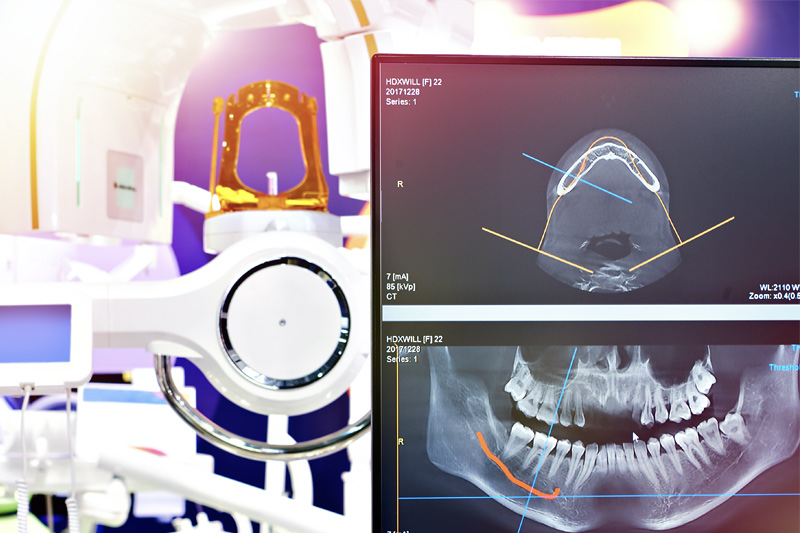
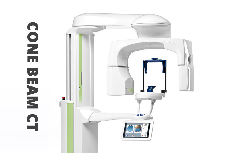
I-CAT CBCT Cone Beam
We use a state of the art I-CAT CBCT Cone Beam 3 dimensional x-ray to find pathological conditions in our patient’s mouth and surrounding areas. We have been amazed at the ability of these images to show periapical periodontal abscesses that do not show up on traditional radiographs. We can also find areas of ischemic bone disease (Cavitation’s) in extraction sites, measure patients air ways to screen for obstructive sleep disorders, and see a 3 dimension image of nerves and roots.
We are able to provide more accurate dimensional views of the skull and jaws. In addition, while CBCT images also provide cross-sectional (bucco-lingual), axial, coronal, sagittal, panoramic and cephalometric views, there is no compromise between the image quality and how safe it is for our patients. There are many more clinical uses available, including dental implant planning, orthodontics, visualization of impacted teeth, airway studies, spinal studies, TMJ analysis, and diagnosis of dental trauma.
This system is increasingly used across the United States since the early 2000’s and is able to meet the ALARA – As Low as Reasonably Achievable radiation protocols (Exposures as low as 4 µSv).